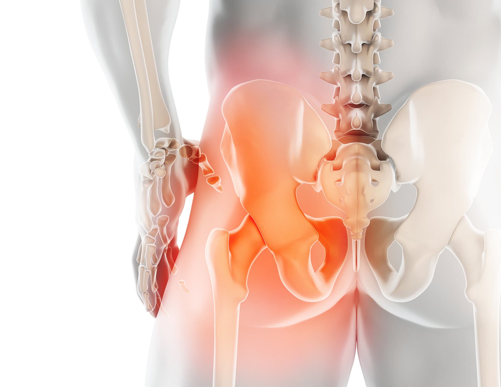
Periacetabular osteotomy
Positioned between arthroscopy and prosthesis, conservative surgery includes corrective procedures for hip malformations: insufficient acetabular coverage (dysplasia), anomalies in the orientation of the femoral neck (coxa valga, coxa vara), rotational disorders.

Principle
Hip impaction surgery involves increasing the acetabular coverage by fixing a bone graft. Periacetabular osteotomy (PAO) involves cutting the pelvis and reorienting the acetabulum. Femoral osteotomy changes the orientation of the neck.
Technique
Periacetabular osteotomy is the reference technique for treating hip dysplasia in young individuals who do not show signs of associated osteoarthritis. However, the frequency of this intervention remains rare in France.
Indications
Periacetabular osteotomy requires precise evaluation of the morphology of the hip joint and the cartilaginous status. Initially, medical treatment is always prioritized, combining weight control, targeted muscle rehabilitation, and a course of anti-inflammatories. Intra-articular injections are also prescribed.
After the failure of complete medical management, the indication for periacetabular osteotomy is re-evaluated in light of the existing cartilaginous lesions, the level of daily disability, and the objectives in terms of recovery.
Operation
Objective
A conservative intervention requires an hour and a half of surgery and five nights of hospitalization. The periacetabular osteotomy is performed according to precise planning aimed at correcting the bone malformation. The goal is to restore a more physiological shape to the hip to delay its joint degradation. Periacetabular osteotomy does not alter the shape of the pelvis and allows for future pregnancies.
Technique
Periacetabular osteotomy is performed under general anesthesia. An incision is made straddling the iliac crest and the top of the thigh. Four osteotomy lines are performed, preserving the posterior column of the iliac bone. These four “around the acetabulum” osteotomies allow detaching the acetabulum from the iliac bone to reorient it in a suitable position. The new positioning of the acetabulum is controlled by images under an image intensifier. Periacetabular osteotomy is most often fixed with 2 screws.
Postoperative Recovery
Walking is gradually resumed, assisted for a few weeks by two crutches. Full activity resumption requires several months, the time for bone healing. Recovery after conservative surgery is longer than after arthroscopy or prosthesis: it is therefore an investment to protect the future of the hip.