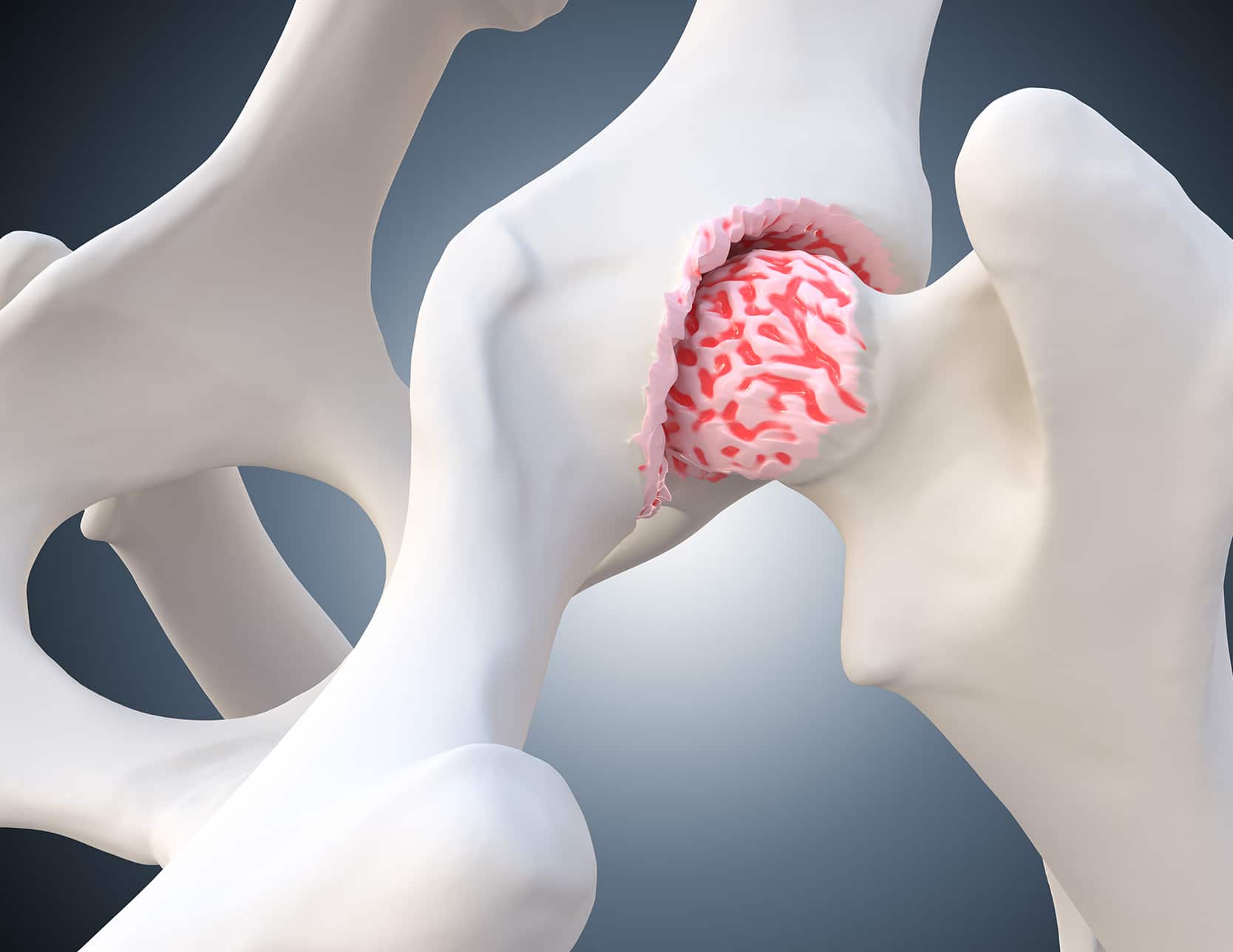
Cartilage Lesions
Also known as chondropathies, cartilage lesions in the hip have multiple causes: high-risk sports activities, trauma or microtraumas, malformations, childhood coxopathy, and a familial predisposition to osteoarthritis. Most commonly, early chondropathy occurs in athletes as a result of hip impingement or dysplasia.
Examination
A lesion of the hip cartilage becomes painful when it is associated with synovitis, a labrum lesion, or subchondral edema. The pain is mechanical in nature, often located in the groin or around the hip, and sometimes radiates to the thigh and knee. Walking distance can be limited, and engaging in sports becomes challenging. Sometimes, cracking sounds in the hip are felt.
The clinical examination looks for joint stiffness and pain during tests for coxopathy. Imaging assessment is used to screen for hip impingement and dysplasia, and to look for narrowing of the joint space on the face and on the Lequesne false profile.
Arthro-CT scanning is the reference examination to identify chondropathy and to specify its depth and extent. MRI is used to explore edema in the subchondral bone.
Treatment
Acetabular chondropathies are common, often associated with hip impingement or dysplasia. If medical treatment is ineffective, hip arthroscopy is considered to correct the cause of impingement, repair the labrum lesion, and resect unstable cartilage lesions. Subchondral perforations are also performed during arthroscopy if the subchondral bone is exposed. In cases of severe dysplasia, a periacetabular osteotomy or prosthesis implantation is discussed.
Femoral chondropathies are rarer and can be associated with osteochondritis of the femoral head, sometimes in a traumatic context. Arthroscopic treatment allows for the resection of unstable cartilage lesions. Osteochondral grafts are exceptionally proposed.
When cartilage lesions are severe and associated with advanced osteoarthritis, treatment relies on prosthesis implantation. In young and athletic individuals, a custom implant, implanted via the anterior approach, is preferred.