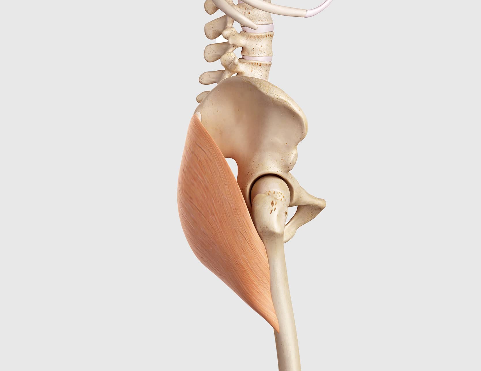
Gluteal tendon repair
Lesions of the gluteal tendons (inflammatory enthesopathy or rupture) are common in women over 60, especially when overweight. Surgical treatment mainly involves the repair of the gluteal tendons combined with the excision of inflammation around the tendons (bursitis).

Natural History
Gluteus medius tendinopathy can progress gradually and lead to a potentially serious tendon rupture. The condition generally begins with a deep inflammation of the tendon, causing lateral pain (trochanteric relief). The infiltration of a cortisone shot around the tendon, often performed by a radiologist under ultrasound control, helps to relieve pain and limping.
Ultrasound and hip MRI also help to specify the extent of the lesions (bursitis, tendinopathy, rupture). At the terminal stage, the rupture can extend to the posterior tendon of the gluteus medius, which ensures the stability of the hip (single-leg support). In the event of a rupture, walking without a cane becomes impossible, and the powerful gluteus medius muscle progresses to permanent fatty degeneration. The rupture of the gluteal tendons is a rare but serious pathology for the stability of the hip.
Medical Treatment of Gluteal Pathology
Fissuring tendinobursitis of the glutes is a condition that is treatable with medical therapy in 95% of cases. It is a long and difficult lesion to treat. Follow-up in sports medicine or rheumatology may be advised. Initial treatment uses pain relievers and anti-inflammatories, gentle rehabilitation, and balneotherapy. Injections in contact with the bursitis should be discussed with your surgeon and must be performed by an experienced doctor under ultrasound control. Finally, shock wave therapy is interesting to stimulate the healing of the damaged tissues.
Tendon Repair
Tendon repair can be performed either by arthroscopy or conventional surgery. The first step is to establish an accurate assessment of the lesions. Tendon rupture is often extensive and associated with a split in the torn tendon. It is then appropriate to resect the poor-quality tendon to find a healthy tendon, then to freshen the bony relief of the major trochanter and fix the tendon with 4 to 6 points using resorbable bone anchors.
Postoperative Recovery
A few hours after the operation, walking can be resumed with partial weight-bearing using two crutches. Crutches are gradually abandoned from 6 weeks, when rehabilitation begins (strengthening work and massages).
Tendon healing is long, results are noticeable at 6 months, but the results are good if the muscle is not atrophic or suffering from fatty degeneration. The tendon repair surgery must be performed before the rupture of the posterior tendon.