Published on 19 November 2025 Hip surgery
Correcting a leg length discrepancy after total hip replacement
After total hip replacement, some patients experience a feeling that one leg is longer or shorter than the other. This imbalance, sometimes causing instability, limping, or lower back pain, may result from a temporary adjustment issue or from an actual change in joint geometry. At Inside the Hip, a highly specialized center in Lyon, 3D planning and custom-made implants are used to eliminate this risk and restore perfect alignment of the pelvis and lower limbs for optimal functional recovery.

3D Planning : a solution to prevent leg length discrepancy
At Inside the Hip , patients scheduled for hip replacement benefit from 3D CT-based preoperative planning followed by the design of a custom-made implant. Thanks to the precision of 3D imaging and custom manufacturing, the risk of leg length discrepancy after surgery is virtually eliminated or extremely low.
Our team is regularly consulted by patients who did not have access to this technique and who have felt a leg length difference since their initial surgery. This sensation often appears immediately after the first postoperative mobilization : an impression that the knees are misaligned, a sense of imbalance, limping, or feeling as though one must “climb a small step” with each stride. Such discrepancies may also cause lumbar or lumboradicular pain, as well as multiple muscular tensions due to compensatory postures.
Possible adjustments after unhappy hip replacement
In some cases, this sensation resolves within a few weeks with rehabilitation and physiotherapy. Consultation with a podiatrist can help by creating a custom plantar orthotic to balance the lower limbs.
However, orthotics alone cannot correct significant leg length discrepancies, nor can they readjust altered muscular and joint tensions resulting from the surgical modification of hip mechanics.
Imaging assessment : a key step in diagnosis
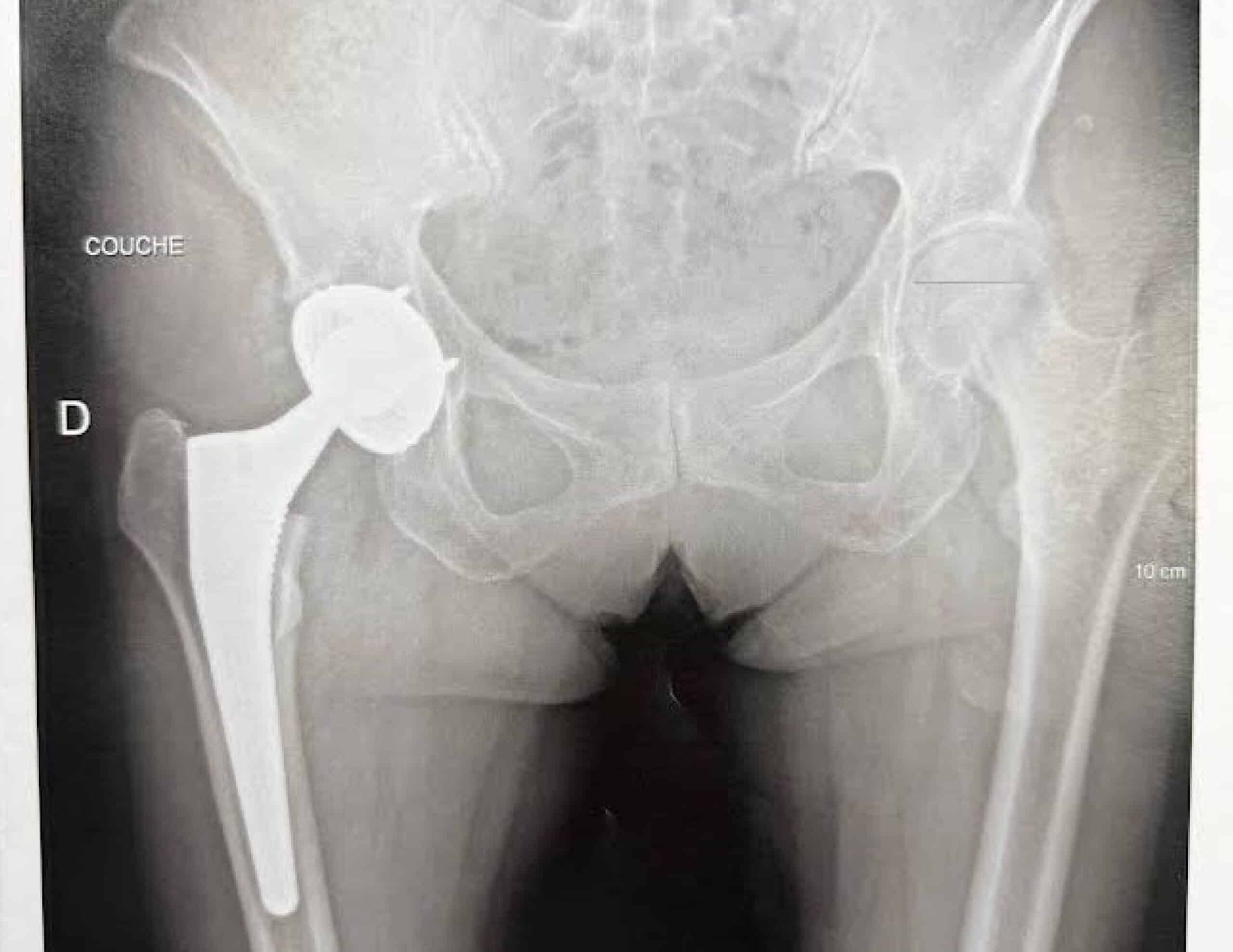
Radiological assessment is essential to quantify the leg length difference. Standing anteroposterior pelvic radiographs, both pre- and postoperatively, should be analyzed.
An EOS full-body standing scan (spine and lower limbs) can measure bone segment lengths and assess the lumbopelvic-femoral balance.
However, the reference examination remains 3D CT imaging, which allows precise geometric analysis.
Unfortunately, many patients did not undergo preoperative CT imaging, making it difficult to obtain accurate measurements of native joint geometry.
When a preoperative CT scan is available, a postoperative scan can perfectly reveal the geometric changes induced by surgery. If not, a bilateral CT scan can compare the operated and non-operated hips to identify asymmetries.
3D CT analysis to identify the cause of discrepancy
3D CT analysis is critical, as the sensation of leg length inequality often combines three interrelated mechanical issues :
- Excessive vertical lengthening (cranio-caudal axis)
- Uncontrolled variation of the medio-lateral offset
- Uncontrolled change in femoral anteversion (axial plane)
Through detailed 3D analysis, it becomes possible to identify cup malposition, femoral stem malalignment, or inaccurate adjustment of soft-tissue tension related to the choice of prosthetic head.
Surgical options to correct leg length discrepancy
Restoring an optimal lumbopelvic-femoral balance should never be neglected.
To achieve this, revision surgery may be considered, depending on the underlying cause:
- Replacement of the prosthetic head only
- Revision of the acetabular cup
- Replacement of the femoral stem
Depending on the case, the revision can be performed through a direct anterior approach, which preserves muscle structures, or a posterior approach, allowing better access to the femur.
In certain complex cases, a custom-made revision implant may be required to restore the hip’s native anatomy and symmetry.
Prevention: the key to long-term balance
Preventing leg length discrepancy relies on a thorough preoperative clinical examination combined with 2D and 3D imaging (particularly preoperative CT).
Choosing a custom-made implant ensures secure and precise adjustment of joint balance, which is essential for a natural gait and full functional recovery.
3D planning and custom implants are especially valuable for young, athletic patients or those with pre-existing spinal conditions, as these cases demand high precision and biomechanical harmony.
